Wall-motion scoring
Score visible LV segments, review WMSI, and describe the observed wall pattern. Coronary territory maps are orientation aids, not patient-specific vessel diagnosis.
Clinically reviewed 25 May 2026 · EchoRapp clinical content
Simple approach
Score each visible LV segment in the views where endocardial motion and systolic thickening are clearest.
| Score | Meaning | Use |
|---|---|---|
| 0 | Not visualized | Segment cannot be scored |
| 1 | Normal | Normal inward motion and thickening |
| 2 | Hypokinetic | Reduced motion or thickening |
| 3 | Akinetic | Absent systolic thickening or motion |
| 4 | Dyskinetic | Systolic outward motion |
| 5 | Aneurysmal | Persistent aneurysmal deformation |
WMSI is the average of the scored segments. A complete 16-segment stage is easiest to compare, but an incomplete stage can still be useful when the count makes missing segments visible.
Describe the observed pattern first: for example, apical abnormality, inferior and inferoseptal abnormality, or abnormal segments 8, 13, 14, 15. Do not let a territory map replace the echo pattern.
Detailed tutorial
WMSI
Segments scored as 0 are not included in the average. A WMSI of 1.00 means all scored segments are normal. Values above 1.00 mean at least one scored segment is abnormal.
In stress echocardiography, compare the same segments across stages. New wall-motion abnormality in a segment that was normal at rest, or worsening in a segment with resting abnormality, is the key ischemia signal. Also review the number, location, and severity of abnormal segments.
Pattern wording
Use wall and segment wording because it is directly supported by the images. A concise pattern is usually enough:
- No abnormal segments scored.
- Apical abnormality.
- Anterior and anteroseptal abnormality.
- Inferior and inferoseptal abnormality.
- Abnormal segments 8, 13, 14, 15.
Typical coronary territory map
Warning
Coronary territory maps are teaching aids. They show common distributions, but individual supply varies with dominance, wraparound LAD, diagonal and marginal branches, prior infarct, bypass grafts, and multivessel disease. Use the map to orient the pattern, not to make a patient-specific vessel diagnosis.
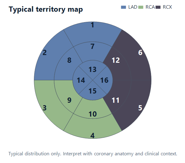
The diagram uses the EchoRapp TTE bullseye orientation: anterior segments are superior, inferior segments are inferior, septal segments are on the left side of the display, and lateral segments are on the right side of the display.
Typical teaching maps often place segments 1, 2, 7, 8, 13, 14, 15, and 16 in the LAD region; segments 3, 4, 9, and 10 in the RCA region; and segments 5, 6, 11, and 12 in the RCX region. This is only a common pattern. Inferior, inferoseptal, inferolateral, anterolateral, and apical supply can vary substantially between patients.
References
- Roberto M. Lang et al. Recommendations for Cardiac Chamber Quantification by Echocardiography in Adults: An Update from the American Society of Echocardiography and the European Association of Cardiovascular Imaging. European Heart Journal 2015;16(3):233–271. View guideline
- Patricia A. Pellikka et al. Guidelines for Performance, Interpretation, and Application of Stress Echocardiography in Ischemic Heart Disease: From the American Society of Echocardiography. J Am Soc Echocardiogr 2020;33(1):1–41. View guideline
- Jose T. Ortiz-Perez et al. Correspondence Between the 17-Segment Model and Coronary Arterial Anatomy Using Contrast-Enhanced Cardiac Magnetic Resonance Imaging. JACC Cardiovasc Imaging 2008;1(3):282–293. View guideline
- Osvaldo Pereztol-Valdes et al. Correspondence between left ventricular 17 myocardial segments and coronary arteries. Eur Heart J 2005;26(24):2637–2643. View guideline